
[18]F-flutemetamol amyloid PET in an MCI patient that later developed AD
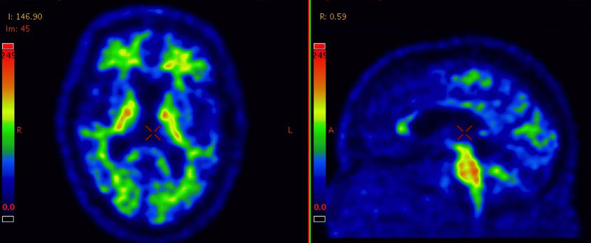
[18]F-flutemetamol amyloid PET in a cognitively healthy elderly
The BioFINDER team is in collaboration with prof Per Wollmer and his team at Lund University and GE Healthcare (USA) measuring the Aβ deposition in the brain of the cognitively healthy elderly and the patients with a mild cognitive impairment. The examination is done using PET imaging and the radio ligand 18F-Flutemetamol (Vizamyl) that now has been approved by the U.S. Food and Drug Administration (FDA) and the European Medical Agency (EMA). All subjects are examined on the same type of scanner (a Philips Gemini TF 16) to avoid technical biases in the results.